
Troponins T & I
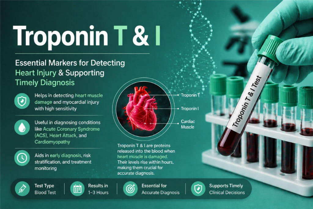
Table of Contents
Definition
Troponin is a regulatory protein complex located on the thin filament of striated muscle tissue. Working alongside tropomyosin, it modulates the calcium-dependent interaction between actin and myosin, which drives muscle contraction.
The complex consists of three distinct subunits:
Troponin C (TnC): Binds calcium ions to initiate contraction. Note: TnC is identical in both cardiac and skeletal muscle, making it useless as a cardiac biomarker.
Troponin I (TnI): The inhibitory subunit that binds to actin to prevent contraction in the absence of calcium.
Troponin T (TnT): The structural subunit that anchors the entire troponin complex to the tropomyosin strand.
Clinical Sinificance
The clinical significance of Trop T and I extends far beyond simply diagnosing a heart attack. In modern medicine, these biomarkers serve as critical diagnostic tools, precise prognostic indicators, and drivers of therapeutic decision-making.
Because high-sensitivity assays can detect minute amounts of myocardial injury, understanding how to interpret these markers within a broader clinical framework is essential for preventing both under-diagnosis and unnecessary, invasive treatments.
1. The Diagnostic Framework: Injury vs. Infarction
The foundational clinical significance of cardiac troponins lies in differentiating between myocardial injury and acute myocardial infarction (AMI). According to the Fourth Universal Definition of Myocardial Infarction, the differentiation relies on a precise combination of biochemical and clinical data:
Myocardial Injury: Documented when at least one cardiac troponin value is above the 99th percentile upper reference limit (URL). The injury is classified as chronic if troponin levels remain elevated and stable (less than a 20% variation) across serial samples, or acute if there is a rising and/or falling pattern.
Myocardial Infarction (Type 1 or Type 2): Documented when there is acute myocardial injury plus clinical evidence of acute myocardial ischemia. This requires at least one of the following:
Symptoms of acute ischemia (e.g., crushing chest pain, dyspnea, radiating left arm pain).
New ischemic ECG changes (e.g., ST-segment elevation, ST-segment depression, or new left bundle branch block).
Development of pathological Q waves on the ECG.
Imaging evidence of new loss of viable myocardium or a new regional wall motion abnormality.
Identification of a coronary thrombus via angiography or autopsy.
2. Differentiating Type 1 vs. Type 2 Myocardial Infarction
When acute troponin elevation meets the criteria for an MI, clinicians look closely at the underlying cause to guide treatment. Troponin kinetics and clinical presentation help categorize the event:
Type 1 MI (Atherothrombotic)
This is the classic heart attack. It is caused by the acute rupture or erosion of an atherosclerotic plaque, leading to thrombus formation and sudden coronary artery occlusion.
Clinical Approach: Requires immediate antiplatelet therapy, anticoagulation, and urgent invasive strategies like cardiac catheterization (percutaneous coronary intervention, or PCI).
Type 2 MI (Ischemic Imbalance)
This occurs secondary to a severe mismatch between myocardial oxygen supply and demand, without acute plaque rupture. Common triggers include severe anemia, profound hypotension, acute respiratory failure, sustained tachyarrhythmias, or severe coronary artery vasospasm.
Clinical Approach: Treatment targets the underlying medical condition (e.g., transfusing blood for severe anemia, or lowering heart rate in tachyarrhythmias) rather than opening an occluded artery.
3. Non-Ischemic Causes of Troponin Elevation
Because troponin is a organ-specific marker (specific to the heart) but not disease-specific, any condition that causes myocardial strain, cellular necrosis, or increased cell membrane permeability can cause an elevation.
Recognizing these non-ischemic etiologies is a core competency for laboratory and clinical teams:
Acute Cardiac Non-Ischemic Conditions
Myocarditis and Pericarditis: Direct viral or autoimmune inflammation of the myocardium damages myocytes, causing significant troponin spikes that often mimic an acute infarction.
Acute Heart Failure Exacerbation: Increased ventricular wall stretch and wall tension lead to subendocardial ischemia and continuous, low-level troponin release.
Takotsubo Cardiomyopathy: Stress-induced cardiomyopathy caused by a massive catecholamine (adrenaline) surge, leading to transient apical ballooning and minor-to-moderate troponin leakage.
Systemic Non-Ischemic Conditions
Pulmonary Embolism (PE): A large blood clot in the lungs causes acute right ventricular strain and pressure overload, leading to right ventricular ischemia. Elevated troponin in a confirmed PE patient is a critical marker of poor prognosis.
Sepsis and Septic Shock: Circulating bacterial endotoxins, severe systemic hypotension, and high metabolic demand cause diffuse myocardial strain and direct cytokine-mediated myocyte injury.
Stroke and Subarachnoid Hemorrhage: Severe central nervous system injury causes an intense autonomic surge, releasing massive amounts of norepinephrine that can trigger focal myocardial necrosis.
4. Prognostic Value and Risk Stratification
Beyond immediate diagnosis, the absolute concentration and baseline trends of Trop T and I hold powerful long-term prognostic weight.
In Acute Coronary Syndromes (ACS)
The magnitude of troponin elevation correlates directly with the size of the myocardial infarct and the risk of mortality. Higher peak values predict an increased risk of developing post-infarct heart failure, cardiogenic shock, and malignant ventricular arrhythmias.
In Asymptomatic and Chronic Populations
With high-sensitivity assays, measurable troponin levels within the “normal” range (but in the upper quartiles) are highly predictive of future adverse outcomes. In patients with stable coronary artery disease, diabetes, or chronic hypertension, steadily rising baseline troponin levels serve as an early warning sign for:
All-cause and cardiovascular mortality.
Future clinical heart failure development.
Subclinical, progressive structural heart disease.
5. Rapid Rule-Out and Rule-In Protocols
The implementation of high-sensitivity troponin assays (hs-cTn) revolutionized emergency department workflows by allowing clinicians to safely discharge low-risk chest pain patients within hours, dramatically reducing hospital overcrowding.
The European Society of Cardiology (ESC) strongly endorses 0-hour/1-hour and 0-hour/2-hour rapid algorithms using validated hs-cTn thresholds:
Rule-Out Strategy: If a patient’s baseline (0-hour) troponin is below the limit of detection, or if it is very low and does not rise after 1 or 2 hours ($\Delta$ troponin is negligible), the patient has a negative predictive value greater than 99% for an AMI and can be safely considered for discharge.
Rule-In Strategy: If the baseline troponin is significantly high, or if the follow-up sample shows an absolute increase (delta) above a pre-set threshold within 1 to 2 hours, the patient is ruled in for further cardiac management.
Methods & Principle
1. The Core Analytical Principle: The Two-Site Sandwich Immunoassay
Almost all automated laboratory platforms utilize a non-competitive, two-site sandwich immunoassay format to measure cardiac troponins. Because troponin proteins are relatively large antigens, they possess multiple distinct regions (epitopes) that can be targeted simultaneously by different antibodies.
Step-by-Step Reaction Mechanics
1.Antigen Capture:Phase 1.
The patient’s sample (serum or plasma) is mixed with the assay reagents. A highly specific capture antibody binds to a unique epitope on the circulating cardiac troponin molecule. This capture antibody is physically anchored to a solid phase—typically paramagnetic microparticles (beads).
2.Sandwich Formation:Phase 2.
A second antibody, the detection antibody, is introduced. This antibody targets a completely different, non-overlapping epitope on the same troponin molecule. The detection antibody is chemically conjugated to a signal-generating label (such as an enzyme or a chemiluminescent molecule). The troponin antigen is now “sandwiched” between the capture and detection antibodies.
3.The Separation Wash:Phase 3.
The testing instrument applies a powerful magnetic field to immobilize the paramagnetic beads holding the antibody-antigen sandwich. The system then performs a vigorous wash cycle to flush away all unbound proteins, matrix elements, and excess detection antibodies. This step is critical to eliminate background noise.
4.Signal Generation and Quantitation:Phase 4.
An activation reagent is introduced to trigger the detection label. The instrument’s internal optical system (a photomultiplier tube) counts the emitted photons or measures light absorbance. Because this is a non-competitive assay, the intensity of the generated signal is directly proportional to the concentration of troponin present in the patient’s sample.
2. Advanced Detection Methodologies
While the underlying sandwich principle remains consistent, different instrument manufacturers utilize distinct biochemical pathways to generate and read the analytical signal.
A. Chemiluminescence Immunoassay (CLIA)
Widely considered the industry standard for high-throughput automated platforms, CLIA utilizes chemical reactions to emit light.
The Reaction: The detection antibody is typically labeled with Acridinium ester or Isoluminol. Upon adding an oxidizing trigger solution (such as hydrogen peroxide in an alkaline medium), an unstable intermediate forms. As this intermediate decays back to its ground state, it flashes light at a specific wavelength (typically ~425 nm).
Clinical Advantage: Exceptional signal-to-noise ratio, enabling the picogram-level detection required for high-sensitivity assays.
B. Electrochemiluminescence Immunoassay (ECLIA)
This is the proprietary pathway utilized exclusively by Roche Diagnostics for their cardiac Troponin T (hs-cTnT) assays.
The Reaction: The capture antibody is biotinylated, binding to streptavidin-coated magnetic microparticles. The detection antibody is labeled with a Ruthenium complex ([Ru(bpy)_3]^2+)
The Mechanics: The reaction occurs on the surface of an electrode. When a voltage is applied, a cyclic electron-transfer reaction between the Ruthenium complex and a co-reactant (Tripropylamine, or TPA) produces an excited state that emits light at 620 nm. The Ruthenium is regenerated in the process, allowing for sustained signal generation.
C. Enzyme-Linked Fluorescent Assay (ELFA)
Frequently used in point-of-care (POC) or smaller benchtop systems.
The Reaction: The detection antibody is conjugated to an enzyme, usually Alkaline Phosphatase (ALP). A fluorogenic substrate (like 4-Methylumbelliferyl phosphate) is introduced. The enzyme cleaves the substrate, producing a fluorescent product that glows under UV light.
3. High-Sensitivity (hs) Assay Engineering: How It Works
To elevate a standard troponin assay to a “high-sensitivity” assay, manufacturers engineered modifications to push the functional detection limits lower by a factor of 10 to 100.
Epitope Protection: Modern assays use combinations of monoclonal antibodies that target the stable, central core of the troponin molecule. This prevents false negatives caused by in-vivo protein degradation or proteolytic cleavage in circulation.
Increased Sample Volume: Adjusting the fluidic sample probes to draw a larger volume of patient serum, packing more troponin molecules into the reaction cuvette.
Mutated Recombinant Antibodies: Genetic engineering of the reagent antibodies to maximize their binding affinity (K_a), ensuring they tightly bind troponin even at trace concentrations (< 2 pg/mL).
4. Pre-Analytical Requirements and Sample Processing
The reliability of a troponin value depends heavily on strict adherence to pre-analytical laboratory protocols.
| Factor | Requirement / Impact |
| Specimen Type | Lithium Heparin Plasma (green-top) or Serum (red/gold-top). Plasma is preferred in emergency medicine because it eliminates the 30-minute clotting wait time, shortening turnaround time (TAT). |
| Centrifugation | Samples must be thoroughly spun (typically 10-15 minutes at 1000-2000 x g). Fibrin microclots remaining in unseparated plasma can plug sample probes or cause false-positive elevations by physically trapping detection antibodies. |
| Hemolysis Interference | Critical Concern: Gross hemolysis causes a significant false decrease in Roche hs-cTnT assays because intracellular protease enzymes released from ruptured red blood cells actively degrade the Troponin T protein. |
| Stability | Separated serum/plasma is stable at room temperature for 8 hours, at 2–8°C for 24 hours, and can be frozen at -20°C or lower for long-term storage. Avoid repeated freeze-thaw cycles. |
Specimen Requirements
To guarantee the clinical validity of high-sensitivity Trop T and I assays, precise adherence to specimen collection and processing guidelines is non-negotiable. Because these assays measure analytes at the picogram level, even minor pre-analytical variances can lead to critically delayed diagnoses or false-positive interventions.
1. Acceptable Sample Matrices: Plasma vs. Serum
While both serum and plasma can be validated on modern chemistry analyzers, Lithium Heparin plasma has become the industry standard for emergency and critical care pathways due to its impact on Turnaround Time (TAT).
A. Lithium Heparin Plasma (Green-Top Tube)
Additive: Lithium Heparin (with or without a plasma separator gel). Heparin accelerates the action of antithrombin, blocking the coagulation cascade and keeping the blood fluid.
Clinical Advantage: Elimination of the mandatory clotting phase. Samples can be centrifuged immediately upon arrival in the lab, saving 30–45 minutes—a crucial factor for meeting the standard <60-minute TAT for cardiac biomarkers.
Key Restriction: Avoid Ammonium Heparin or Sodium Heparin tubes, as these can interfere with specific enzymatic or electrochemical detection methods on certain platforms.
B. Serum (Red-Top or Gold-Top SST Tube)
Additive: Clot activator with or without a hydrophilic polymer gel matrix (Serum Separator Tube).
Processing Deficit: Must be allowed to clot completely for 30–60 minutes at room temperature (20-25 C) before centrifugation.
Incomplete Clotting Risk: If a serum sample is spun too early, latent fibrin strands will continue to form in the separated serum. These micro-fibrin clots can obstruct analyzer aspiration probes or coat the reaction cuvette surface, causing erratic fluid delivery and falsely elevated results due to non-specific binding.
2. Tube Inversion and Centrifugation Parameters
Proper mixing and centrifugation ensure clean phase separation and eliminate particulate matter that can distort light-path measurements.
Inversion: Immediately after phlebotomy, both plasma and serum tubes must be gently inverted 5 to 8 times. Vigorous shaking causes mechanical hemolysis, while inadequate mixing in plasma tubes leads to partial micro-clotting.
Centrifugation Speed & Time:
Standard Swing-Bucket Rotor: Centrifuge at 1,000-2,000 x g for 10 to 15 minutes.
Stat Centrifuges: Fixed-angle, high-speed stat processing may utilize 3,000 x g or higher for 5 minutes, provided the tube manufacturer has validated the gel barrier stability at higher forces.
Temperature: Centrifugation should occur at room temperature (20-25 C). Refrigerated centrifugation (4 C) is unnecessary and can cause gel barrier hardening, preventing proper separation.
3. Strict Pre-Analytical Interferences
The primary job of a lab specialist running troponins is evaluating sample integrity. Three main endogenous interferences can alter the true values of Trop T and I:
A. Hemolysis (The Most Critical Interference)
Hemolysis introduces intracellular contents into the sample matrix, creating direct chemical interference:
The Roche hs-cTnT Vulnerability: Grossly hemolyzed specimens cause a significant false decrease in cardiac Troponin T values on Roche platforms. Intracellular proteolytic enzymes released from ruptured erythrocytes actively digest the core troponin T protein fragments, rendering them unrecognizable by the reagent antibodies.
Troponin I Variance: Hemolysis impact on Troponin I is highly platform-dependent; it can cause either a false decrease or an artificial elevation depending on whether the vendor’s antibody epitopes are blocked or cross-reactive with free hemoglobin.
Lab Policy: Samples exceeding the manufacturer’s validated Hemolysis Index (H-Index) threshold must be rejected, and a redraw must be requested immediately.
B. Lipemia
Extremely turbid, milky samples (due to elevated triglycerides or chylomicrons) cause physical light scattering during the optical phase of immunoassays.
Remedy: If lipemia indices exceed limits, samples must be cleared using a high-speed micro-centrifuge or an airfuge to separate the lipid layer before testing the underlying clear fraction.
C. Icterus
High concentrations of bilirubin can absorb light at wavelengths close to those used by chemiluminescent or enzymatic triggers. Most modern high-sensitivity assays are highly resistant to icterus up to a Bilirubin Index of approximately $20\text{–}30 \text{ mg/dL}$, but local platform limits must be carefully monitored via internal quality control.
4. Specimen Stability and Storage
When serial sampling is required, or when a clinician requests a reflex add-on test, understanding analyte stability is vital.
| Storage Temperature | Maximum Validated Stability | Clinical & Analytical Context |
| Room Temperature (20-25 C) | 8 Hours | Samples must be capped to prevent evaporation. If analysis is delayed past 8 hours, move to refrigeration. |
| Refrigerated (2-8 C) | 24 to 48 Hours | Ideal window for checking delta values or running add-on confirmations. Matrix degradation accelerates past 48 hours. |
| Frozen (-20 C or lower) | up to 12 Months | For long-term clinical trials or research biobanking. Crucial Rule: Samples must be thawed completely, mixed well, and never subjected to repeated freeze-thaw cycles, which break apart the troponin protein structure. |
Reference Ranges & Clinical Interpretations
The clinical utility of high-sensitivity Trop T and I relies entirely on the precise application of reference ranges and the calculation of kinetic changes (deltas). Because these modern assays can detect minute amounts of troponin in healthy individuals, establishing a clear line between “normal physiological presence” and “pathological myocardial injury” is essential.
1. Defining the Reference Range: The 99th Percentile URL
The universal standard for establishing a normal reference range for cardiac troponin is the 99th percentile Upper Reference Limit (URL).
Definition: The concentration below which 99% of a rigorously screened, ostensibly healthy reference population falls.
The Clinical Threshold: Any value above the 99th percentile URL signifies myocardial injury.
Why Reference Ranges Cannot Be Standardized
Unlike other routine chemistry analytes (e.g., glucose or creatinine), there is no single reference range for troponin.
For hs-cTnT (Roche): The assay is globally standardized because one manufacturer holds the patent. The standard 99th percentile URL is 14 ng/L (or pg/mL}).
For hs-cTnI (Multi-vendor): Assays manufactured by different companies (Abbott, Beckman Coulter, Siemens, Alere) target different epitopes of the Troponin I molecule. Consequently, their 99th percentile URLs vary significantly—ranging from 15 ng/L to >50ng/L depending on the platform.
Critical Laboratory Rule: A laboratory must strictly utilize the specific 99th percentile URL validated for their specific instrument model and reagent lot.
2. Sex-Specific Cutoffs in High-Sensitivity Assays
One of the most significant advancements of high-sensitivity assays is the requirement for sex-specific reference ranges. Men generally possess greater cardiac muscle mass than women, resulting in higher physiological troponin turnover.
If a laboratory applies a single, blended reference range to all patients, it introduces significant diagnostic bias:
Under-diagnosing Women: A blended cutoff will be too high for women, causing clinicians to miss subclinical or evolving myocardial infarctions in female patients presenting with atypical symptoms.
Over-diagnosing Men: A blended cutoff may be too low for men, leading to unnecessary cardiac catheterizations for minor, non-ischemic elevations.
Typical Sex-Specific Thresholds (Example: Abbott Alinity hs-cTnI)
Female 99th Percentile URL: 16 ng/L
Male 99th Percentile URL: 34 ng/L
3. Clinical Interpretation: Kinetic Deltas
A single elevated troponin result indicates myocardial injury, but it does not tell you if the injury is an acute event (like a heart attack) or a chronic condition (like structural heart disease or renal failure). To differentiate, laboratories and emergency departments calculate the kinetic delta —the absolute or percentage change between serial samples drawn 1, 2, or 3 hours apart.
Interpretation Framework
1. Acute Myocardial Injury (Rising or Falling Pattern)
Criteria: The baseline or follow-up troponin exceeds the 99th percentile URL, and the change between the samples (Delta) exceeds a pre-determined clinical threshold (e.g., an absolute change of >3-5 ng/L within 1–2 hours, or a > 20% change at 3 hours).
Clinical Meanings: Acute Myocardial Infarction (Type 1 or 2), acute myocarditis, acute heart failure decompression, or a massive pulmonary embolism.
2. Chronic Myocardial Injury (Stable Pattern)
Criteria: Troponin values are persistently elevated above the 99th percentile URL, but remain stable, varying by less than the clinical delta (typically < 20% variation) across serial draws.
Clinical Meanings: Chronic kidney disease (CKD), structural heart disease (e.g., severe aortic stenosis), chronic hypertensive heart disease, or long-standing stable cardiomyopathy.
4. Critical Interpretation Grids for the Medical Laboratory
To assist in rapid clinical decision-making, laboratories often categorize high-sensitivity troponin values into specific interpretation tiers based on the baseline concentration:
| Value Tier | Clinical Interpretation | Action Protocol |
Below Limit of Detection (LoD) (e.g., < 2-3 ng/L | Highly improbable acute ischemia. If chest pain onset occurred >3 hours prior to phlebotomy, this has a >99% Negative Predictive Value (NPV) for AMI. | Rule-Out Complete: Safe for discharge regarding AMI, pending clinical scoring (e.g., HEART score). |
Normal Range (LoD up to 99th Percentile) | No current evidence of biochemical myocardial necrosis. | Observe and Re-test: If chest pain is highly recent (<3 hours), a second sample must be evaluated at 1–2 hours to monitor for an early kinetic rise. |
Mild to Moderate Elevation (99th Percentile up to 3x URL) | Myocardial injury present. Can be due to early-stage AMI or chronic/systemic conditions (CKD, heart failure, sepsis, pulmonary embolism). | Calculate Kinetic Delta: Run a follow-up sample at 1–2 hours. A significant change confirms an acute injury; stable values point toward chronic etiology. |
Marked Elevation / Critical Alert ( >3x to 10x+ the URL) | Severe, acute myocardial necrosis. Highly specific for an acute atherothrombotic MI (Type 1) or massive acute cardiac insult. | Immediate Rule-In: Critical values are called immediately to the physician. Patient is prioritized for urgent cardiology assessment or coronary intervention. |
Quick Stats
| Feature | Details | Critical Lab Insights |
| Test Type | Organ-Specific Cardiac Biomarker | The gold standard indicator for myocardial necrosis. It is highly specific to cardiac muscle tissue but not disease-specific, meaning it flags heart damage but not the underlying cause. |
| Sample Type | Lithium Heparin Plasma or Serum | Lithium Heparin (Green Top) is preferred in emergency settings to bypass the 30–60 minute clotting phase required for Serum (Red/Gold Top), drastically improving Turnaround Time (TAT). |
| Detection Method | Two-Site Sandwich Immunoassay | Utilizes a non-competitive format where the troponin antigen is “sandwiched” between a solid-phase capture antibody and a labeled detection antibody. Signal intensity is directly proportional to concentration. |
| Primary Metric | Nanograms per Liter (ng/L) or pg/mL | Expressed in whole numbers (e.g., 14 ng/L instead of 0.014 µg/L). High-sensitivity assays measure down to picogram levels, identifying trace myocardial leaks. |
| Fasting Required? | No | Non-fasting. However, gross lipemia or severe icterus can cause physical light scattering or optical absorbance shifts depending on the analyzer’s light path. |
| Standardization | cTnT: Roche Patented / Standardized cTnI: Multi-Vendor (Non-Standardized) | cTnT has a single global manufacturer and uniform cutoff. cTnI assays are produced by multiple companies targeting different epitopes; numerical results and cutoffs cannot be used interchangeably between different platforms. |
| Turnaround Time | 30–60 Minutes | Critical emergency marker. The laboratory’s goal is a TAT of less than 60 minutes from vein to brain to facilitate rapid rule-out or rule-in clinical pathways in the emergency department. |
| Biological Half-life | ~2 Hours (In-vivo Clearance) | While the free circulating half-life is short, continuous leakage from the structurally bound myofibrillar pool causes serum levels to remain elevated for 5–10 days (cTnI) or 10–14 days (cTnT) post-infarct. |
| Normal Range | Below the 99th Percentile URL | The upper reference limit is sex-specific. Men have higher normal baseline cutoffs due to greater cardiac mass; using a blended cutoff under-diagnoses myocardial infarctions in female patients. |
| Kinetic Delta (Delta) | Platform-Dependent Change Baseline | A rising or falling kinetic delta (e.g., >20% or absolute changes of >3-5 ng/L within 1–2 hours distinguishes acute myocardial injury from chronic, stable elevations. |
| Key Limitation | Pre-Analytical Interferences | Gross hemolysis causes a false decrease in Roche hs-cTnT assays due to red cell protease degradation. High-dose Biotin intake causes a false decrease by competing with the streptavidin-biotin capture system. |
FAQs
Q1: What is the main clinical difference between Troponin T and Troponin I?
Answer: The primary clinical difference lies in their tissue specificity and assay standardization. Cardiac Troponin I (cTnI) is absolutely specific to cardiac tissue and is never expressed by skeletal muscle. Cardiac Troponin T (cTnT) is also highly cardiac-specific, but trace amounts can be re-expressed by regenerating skeletal muscle in chronic diseases like polymyositis or End-Stage Renal Disease (ESRD). Additionally, cTnT is globally standardized by a single manufacturer (Roche), whereas cTnI assays are produced by multiple vendors with varying numerical cutoffs and no global standardization.
Q2: Why does gross hemolysis cause a false decrease in Troponin T assays?
Answer: Gross hemolysis releases intracellular components from ruptured red blood cells directly into the sample matrix. Among these components are specific intracellular protease enzymes. In Roche high-sensitivity Troponin T (hs-cTnT) assays, these proteases actively degrade the core amino acid sequences of the circulating Troponin T protein fragments. Because the reagent antibodies can no longer recognize the degraded epitopes, the analyzer detects a falsely decreased signal, which can dangerously mask an active myocardial infarction.
Q3: How does high-dose Biotin consumption interfere with Troponin results?
Answer: Most modern automated troponin immunoassays utilize a high-affinity biotin-streptavidin capture mechanism to immobilize the antibody-antigen sandwich onto paramagnetic particles. If a patient consumes mega-doses of Biotin (common in hair, skin, and nail supplements), the free biotin in their plasma outcompetes the reagent biotin for the binding sites on the streptavidin-coated beads. In sandwich immunoassays, this competitive interference prevents the sandwich from binding to the solid phase, causing it to be washed away, resulting in a falsely decreased (false-negative) troponin result.
Q4: What constitutes a significant kinetic delta (Delta) for high-sensitivity troponin?
Answer: A significant kinetic delta depends on the specific assay manufacturer and the time interval between blood draws (1, 2, or 3 hours). Generally, for rapid 0-hour/1-hour or 0-hour/2-hour protocols, an absolute change of >3-5 ng/L is considered a significant acute delta. For traditional 3-hour pathways, a relative percentage change of >20% above the baseline value is used. A significant delta confirms acute myocardial injury (e.g., AMI), while a stable troponin value (below the delta threshold) points toward chronic myocardial injury (e.g., CKD or chronic heart failure).
Q5: Why are sex-specific cutoffs mandatory for high-sensitivity troponin assays?
Answer: High-sensitivity troponin assays (hs-cTn) are precise enough to measure baseline troponin levels in healthy individuals. Because biological men typically have a larger left ventricular muscle mass than women, they experience higher physiological myocyte turnover, resulting in higher normal baseline troponin levels. If a lab uses a single “blended” reference range, the cutoff becomes too high for women—leading to the under-diagnosis of myocardial infarctions in female patients—and too low for men, causing false positives.
Q6: Can a patient have an elevated troponin without having a heart attack?
Answer: Yes. Troponin is an organ-specific marker (indicating cardiac damage), but it is not disease-specific. Any condition that causes myocardial strain, cellular ischemia, inflammatory necrosis, or increased cell wall permeability can cause troponin to leak into circulation. Common non-ischemic causes include chronic kidney disease (CKD), severe sepsis, acute pulmonary embolism (PE), myocarditis, acute heart failure exacerbation, and extreme strenuous exercise (like running a marathon).