
D-Dimers
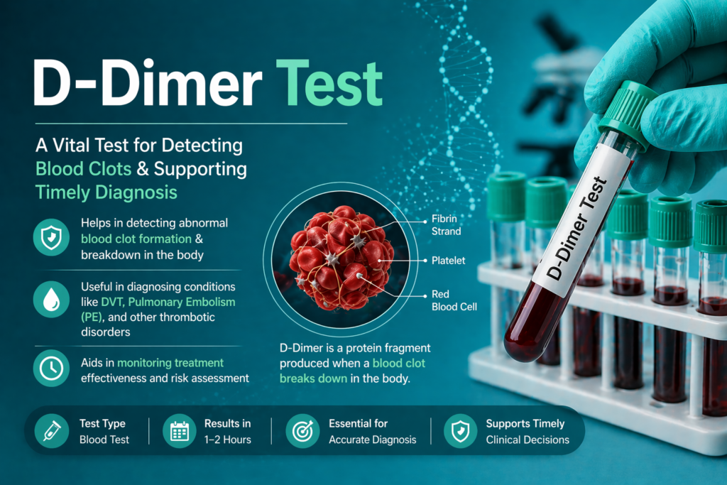
Table of Contents
Definition
D-dimer is a fibrin degradation product (FDP), a small protein fragment present in the blood after a blood clot is degraded by fibrinolysis. It contains two cross-linked D fragments of the fibrin protein.
The Formation Process
Thrombin Generation: Thrombin converts soluble fibrinogen into insoluble fibrin monomers.
Cross-linking: Factor XIIIa cross-links these monomers at the “D” domains to create a stable fibrin mesh.
Fibrinolysis: To prevent vessel occlusion, the enzyme plasmin cleaves the fibrin mesh.
Release: The specific cleavage of cross-linked fibrin (not fibrinogen) releases D-dimer molecules into the circulation.
Clinical Significance
In the clinical laboratory, the D-dimer test is rarely used for “diagnosis” in the traditional sense. Instead, its true clinical power lies in its high sensitivity and high Negative Predictive Value (NPV). For a medical laboratory scientist, understanding the clinical significance means knowing when a result provides a green light for clinicians to stop testing or a red flag to investigate further.
1. Ruling Out Venous Thromboembolism (VTE)
The most significant clinical use of D-dimer is to exclude Deep Vein Thrombosis (DVT) and Pulmonary Embolism (PE).
The Power of “Negative”: If a patient has a low clinical probability (based on a Wells Score or Geneva Score) and a D-dimer result below the laboratory cutoff (typically <500 ng/mL FEU), the probability that they have a clot is nearly zero.
Avoiding Over-Imaging: By confidently ruling out VTE with a blood test, clinicians can avoid expensive and potentially harmful imaging like Computed Tomography Angiography (CTA) or ultrasound, which involve radiation and contrast dyes.
2. Diagnosis and Monitoring of DIC
Disseminated Intravascular Coagulation (DIC) is a life-threatening systemic process where clotting and bleeding occur simultaneously.
Consumption Coagulopathy: In DIC, thrombin is generated throughout the entire vascular system. This leads to massive fibrin formation followed by intense secondary fibrinolysis.
The Trend Matters: In the lab, you will see D-dimer levels that are often 10x to 100x the reference limit. Monitoring the trend of D-dimer (alongside Fibrinogen and Platelet counts) is vital to see if the patient is responding to treatment or if the coagulopathy is worsening.
3. Pregnancy and Preeclampsia
Managing D-dimer in pregnancy is a major clinical challenge.
Normal Pregnancy: D-dimer levels naturally rise throughout each trimester due to the physiological changes in the coagulation system.
Complications: Significantly higher-than-expected D-dimer levels can be an early indicator of Preeclampsia or Placental Abruption. However, because the baseline is already high, labs must often use pregnancy-specific reference intervals to avoid constant false positives.
4. Cardiovascular Health and Aortic Dissection
While not the primary diagnostic tool, an extremely elevated D-dimer in a patient with “tearing” chest pain can increase the suspicion of an Acute Aortic Dissection (AAD). Since an AAD involves a tear in the aortic wall where blood enters and clots, the subsequent fibrinolysis releases D-dimer.
5. Malignancy and Chronic Inflammation
Cancer cells can express tissue factor and other procoagulants. Clinically, a persistently elevated D-dimer in a patient without an obvious clot can sometimes be a “herald” sign of an underlying, undiagnosed malignancy.
Summary Table: Clinical Interpretation
| D-Dimer Level | Typical Clinical Interpretation |
| Negative (< Cutoff) | High confidence in excluding DVT/PE in low-risk patients. |
| Mildly Elevated | Non-specific; could be age, pregnancy, or minor injury. |
| Moderately Elevated | May suggest VTE, recent surgery, or systemic inflammation. |
| Markedly Elevated | Strong indicator of DIC, massive PE, or severe trauma. |
Methods & Principle
For a medical laboratory professional, the “method” is the platform, but the “principle” is the specific biochemical interaction that ensures the test is measuring cross-linked fibrin and not just fibrinogen.
Below is a detailed breakdown of the methodologies and scientific principles currently used in diagnostic laboratories.
1. Automated Latex-Enhanced Immunoturbidimetry
This is the “workhorse” method in most clinical labs (e.g., Sysmex, Stago, Siemens). It balances high sensitivity with a rapid Turnaround Time (TAT), often under 15 minutes.
Principle: Particle-Enhanced Light Scattering.
The Reagent: Contains sub-microscopic polystyrene latex particles. These particles are chemically coated (sensitized) with monoclonal antibodies (mAbs) specifically targeting the neo-epitope of the D-dimer domain.
The Reaction: When patient plasma is mixed with the reagent, D-dimer molecules act as “multivalent antigens.” One D-dimer molecule can bind to antibodies on two different latex particles, creating a bridge. This leads to the formation of large lattice-like aggregates (agglutination).
Measurement: As these aggregates form, the solution’s turbidity increases. The analyzer measures the decrease in light transmission (Turbidimetry) or the increase in light scattering (Nephelometry). The rate of change in absorbance is proportional to the concentration of D-dimer in the sample.
2. Enzyme-Linked Immunofluorescence Assay (ELFA)
Commonly performed on the bioMérieux VIDAS platform, this is often used as a reference method due to its exceptional sensitivity.
Principle: Two-Step “Sandwich” Fluorescence.
Solid Phase: The Solid Phase Receptacle (SPR)—a tip-like device—serves as both the pipetting device and the solid phase coated with anti-D-dimer antibodies.
Mechanism:
Capture: The sample is cycled in and out of the SPR; D-dimer binds to the interior walls.
Labeling: An enzyme-labeled antibody (alkaline phosphatase) is added, forming the “sandwich.”
Detection: A fluorescent substrate (4-Methylumbelliferyl phosphate) is introduced. The enzyme catalyzes the substrate into a fluorescent product.
Result: The intensity of the fluorescence is measured at 450 nm. This method is highly resistant to interference from hemolysis or icterus because it uses fluorescence rather than simple light transmission.
3. Chemiluminescence Enzyme Immunoassay (CLIA)
Used in high-end, fully automated systems like the Werfen ACL TOP or various immunoassay tracks.
- Principle: Magnetic Microparticle Capture & Light Emission.
Mechanism: D-dimer is captured by monoclonal antibodies attached to paramagnetic microparticles. A second antibody, labeled with a chemiluminescent marker (like acridinium ester), is added.
Detection: A “trigger” solution is added, causing a chemical reaction that releases a flash of light.
Advantage: CLIA has a very broad linear range (the ability to measure extremely high DIC levels and very low rule-out levels without manual dilution).
4. Quantum Dot Fluorescence Immunochromatography
An emerging technology used in advanced Point-of-Care (POC) and rapid testing modules.
Principle: Nanocrystal Lateral Flow.
Mechanism: Uses “Quantum Dots” (semiconductor nanocrystals) instead of traditional gold or dye markers. When UV light hits these dots, they emit intense, stable fluorescence.
Execution: The sample moves across a nitrocellulose membrane via capillary action. D-dimer is captured in a “Test Line” of antibodies. The Quantum Dots provide a much stronger signal than older POC tests, allowing for quantitative results that rival central lab analyzers.
Technical Summary for Lab Professionals
| Feature | Immunoturbidimetric | ELFA (VIDAS) | CLIA |
| Detection Mode | Absorbance / Turbidity | Fluorescence | Chemiluminescence |
| Throughput | Very High (Random Access) | Medium | High |
| Sensitivity | High | Very High | Extremely High |
| Sample Prep | Citrated Plasma (PPP) | Citrated Plasma | Citrated Plasma |
| Key Limitation | Sensitive to HIL* | Batch-dependent speed | High reagent cost |
*HIL: Hemolysis, Icterus, Lipemia.
The “Neo-Epitope” Principle
Regardless of the method, the most critical scientific principle is the use of 8D3 or DD3 monoclonal antibodies. These antibodies only recognize the “D-D” cross-link created by Factor XIII. They do not react with Fibrinogen or Fibrinogen Degradation Products (FDPs). This specificity is what allows us to distinguish between simple fibrinogenolysis and true clot breakdown.
Specimen Requirements
In the clinical laboratory, the D-dimer assay is highly sensitive to pre-analytical variables. Because the test measures the end product of the coagulation cascade, any activation of the clotting process outside the body (in vitro) will lead to falsely elevated results and potential clinical mismanagement.
Below are the rigorous specimen requirements necessary to ensure diagnostic accuracy.
1. Primary Collection Details
Proper collection is the first line of defense against erroneous results.
Tube Type: 3.2% Sodium Citrate (Light Blue Top).
Note: 3.8% citrate is sometimes used, but 3.2% is the international standard for coagulation testing. Using the wrong concentration can affect the ionized calcium levels during testing.
Draw Volume: The tube must be filled to the etched fill line.
The 9:1 Ratio: Coagulation tubes contain a fixed amount of liquid anticoagulant. A ratio of 9 parts blood to 1 part anticoagulant is mandatory.
Under-filling: Leads to excess citrate, which binds the calcium in the test reagents, causing falsely prolonged times or inaccurate D-dimer quantification.
Order of Draw: The blue top must be drawn before other additive tubes (like Heparin or EDTA) to prevent cross-contamination of anticoagulants. If using a butterfly needle, a “discard tube” must be used first to prime the tubing and remove air, ensuring the blue top reaches the proper volume.
2. Specimen Handling and Processing
Once collected, the sample must be handled with care to maintain the integrity of the proteins.
Inversion: Gently invert the tube 3 to 4 times immediately after collection. Do not shake, as vigorous agitation causes hemolysis and premature platelet activation.
Centrifugation (The Gold Standard): * Samples must be processed to produce Platelet-Poor Plasma (PPP).
Requirement: Platelet count must be <10 x 10⁹/L.
Procedure: Typically centrifuged at 1500–2000 g for 15 minutes (or 10 minutes in a high-speed “Stat” centrifuge).
Why PPP? Platelets contain Factor XIII and other procoagulants. If they remain in the plasma, they can release intracellular contents or interfere with the optical readings of automated analyzers.
3. Stability and Storage
D-dimer is relatively stable compared to Factor VIII or Protein C, but it still follows strict timelines.
| Storage Condition | Temperature | Stability Duration |
| Room Temperature | 18–25°C | Up to 4–8 hours (capped) |
| Refrigerated | 2–8°C | Up to 24 hours |
| Frozen (Plasma only) | -20°C | Up to 1 month |
| Long-term Frozen | -70°C | Up to 6 months |
Important: If the test cannot be performed within 4 hours, the plasma should be separated (aliqouted) and frozen. Never freeze whole blood, as the red cells will lyse.
4. Rejection Criteria (When to ask for a Redraw)
As a lab professional, you must be prepared to reject specimens that compromise patient safety.
Hemolysis: Hemoglobin interferes with the light-scattering principles used in immunoturbidimetric assays.
Clotted Specimen: If a micro-clot is present in the tube, the D-dimer has already been “consumed” or falsely generated within the tube. The result will not reflect the patient’s actual physiological state.
Lipemia/Icterus: Excessive lipids or bilirubin can interfere with optical detection. While some modern analyzers can “blank” for this, extreme cases require an alternative method (like ELFA).
Incorrect Anticoagulant: EDTA (Purple top) or Heparin (Green top) tubes are strictly prohibited for D-dimer testing as they interfere with the biochemistry of the assay.
Reference Ranges & Clinical Interpretations
For medical laboratory professionals, the interpretation of D-dimer results is more complex than a simple “High” or “Low” reading. Because D-dimer levels naturally fluctuate with age and physiological status, the laboratory must provide clear guidance to prevent the over-diagnosis of thromboembolism.
1. The Standard Reference Limit
The most widely accepted “rule-out” threshold for Venous Thromboembolism (VTE) in the general population is:
Standard Cut-off: < 500 ng/mL FEU (Fibrinogen Equivalent Units)
Alternative Cut-off: < 250 ng/mL DDU (D-Dimer Units)
Critical Warning: Always check your analyzer’s reporting units. Reporting a result of 400 ng/mL as “Normal” when your lab uses DDU (where the limit is 250) could lead to a missed diagnosis.
2. Age-Adjusted D-Dimer Cut-offs
As humans age, fibrinogen turnover increases even in the absence of disease. Using a static 500 ng/mL cut-off for elderly patients results in a high number of false positives.
The Formula: For patients > 50 years old, use:
Age x 10 ng/mL FEUExample: For an 80-year-old patient, the adjusted cut-off is 800 ng/mL.
Clinical Impact: Implementing age-adjusted cut-offs increases the specificity of the test without significantly compromising safety, reducing unnecessary imaging in older populations.
3. Clinical Interpretation of Results
Negative Result (< Cut-off)
Interpretation: High Negative Predictive Value (NPV).
Action: In patients with a Low to Intermediate clinical probability (Wells Score), VTE is effectively ruled out. No further imaging is typically required.
Caveat: If the clinical suspicion is “High,” a negative D-dimer is insufficient to rule out a clot; imaging is still mandatory.
Mildly to Moderately Elevated (Cut-off to 2,000 ng/mL)
Interpretation: Non-specific elevation.
Possible Causes: * Recent trauma or surgery (within the last 14 days).
Active malignancy.
Pregnancy (D-dimer increases by trimester).
Inflammatory conditions (Rheumatoid arthritis, infections).
Acute Coronary Syndrome (ACS).
Markedly Elevated (> 4,000 ng/mL or > 8x Upper Limit)
Interpretation: High probability of significant fibrinolysis.
Possible Causes:
Disseminated Intravascular Coagulation (DIC): Usually accompanied by low platelets and prolonged PT/APTT.
Massive Pulmonary Embolism.
Aortic Dissection.
Severe Sepsis.
4. D-Dimer Patterns in Specific Conditions
Disseminated Intravascular Coagulation (DIC)
In the ISTH (International Society on Thrombosis and Haemostasis) scoring system for DIC, the D-dimer is a “Strong Marker.”
No increase: 0 points
Moderate increase: 2 points
Strong increase: 3 points
Combined with decreasing fibrinogen and rising PT, this confirms a DIC diagnosis.
Pregnancy-Specific Intervals
Standard cut-offs are often useless in the third trimester. Research suggests the following “normal” elevations:
1st Trimester: 0–950 ng/mL FEU
2nd Trimester: 300–1500 ng/mL FEU
3rd Trimester: 400–3100 ng/mL FEU
5. Summary Interpretation Table
| Result Level | Probability of VTE | Common Clinical Follow-up |
| < 500 ng/mL | < 1% (Very Low) | Discharge / Investigate other causes |
| 500–1000 ng/mL | Low / Moderate | Clinical correlation; potential Doppler US |
| 1000–4000 ng/mL | Moderate / High | Diagnostic Imaging (CTPA or Ultrasound) |
| > 4000 ng/mL | Very High | Urgent imaging; check for DIC or Sepsis |
6. The “Hook Effect” in Laboratory Testing
In extremely rare cases of massive D-dimer presence (e.g., severe DIC), an “Antigen Excess” or Hook Effect may occur in immunoturbidimetric assays. The antibodies become saturated with antigen, preventing the formation of lattices, which can lead to a falsely lower result.
Laboratory Tip: If a result does not match the clinical picture (e.g., patient is visibly clotting but D-dimer is moderate), perform a 1:10 dilution with saline to check for linearity.
Quick Stats
| Feature | Details | Critical Lab Insights |
| Test Type | Coagulation / Special Chemistry | A marker of secondary fibrinolysis. It specifically detects cross-linked fibrin fragments, not fibrinogen degradation products (FDPs). |
| Sample Type | Platelet-Poor Plasma (PPP) | 3.2% Sodium Citrate (Light Blue Top). Must be centrifuged at 1500–2000g for 15 mins to ensure a platelet count <10 x 10^9/L. |
| Primary Metric | FEU (ng/mL) or DDU (ng/mL) | FEU (Fibrinogen Equivalent Units) is the most common. Conversion: 1.0 yu/mL FEU = 0.5 yu/mL DDU}. Confusing these leads to major diagnostic errors. |
| Fasting Required? | No | Diet does not impact fibrinolysis. However, lipemic samples can interfere with optical-based (turbidimetric) detection systems. |
| Standardization | Monoclonal Antibody Specificity | Results are standardized using mAbs (e.g., 8D3 or DD3) that only bind to the neo-epitope formed after Factor XIII cross-links fibrin. |
| Turnaround Time | 15–30 Minutes | Usually handled as a STAT request in suspected Pulmonary Embolism (PE) or DIC cases to facilitate immediate clinical decisions. |
| Biological Half-life | ~8 Hours | D-dimer is cleared by the liver and kidneys. Levels drop quickly once the thrombotic event is resolved and fibrinolysis ceases. |
| Rule-Out Cut-off | < 500 ng/mL FEU | In patients with low clinical probability, a result below this threshold has a 99% Negative Predictive Value (NPV) for ruling out DVT/PE. |
| Critical Values | > 4,000 ng/mL FEU | Extremely high values are highly suggestive of DIC, massive pulmonary embolism, or severe systemic inflammation (e.g., Sepsis). |
| Key Limitation | Low Specificity | Levels naturally rise with age, pregnancy, surgery, and malignancy. A positive result is not diagnostic of a clot on its own. |
FAQs
1. Why can’t we use a D-dimer test to “diagnose” a blood clot?
Answer: The D-dimer test has high sensitivity but low specificity.
While almost everyone with a clot will have an elevated D-dimer, many people without a clot will also have an elevated result. Because fibrin is formed and broken down during wound healing, inflammation, infection, and pregnancy, a “positive” result simply means there is fibrinolysis occurring somewhere in the body. It does not pinpoint the location or the cause (e.g., DVT vs. a recent bruise). Therefore, it is a rule-out tool, not a rule-in tool.
2. What is the difference between D-Dimer Units (DDU) and Fibrinogen Equivalent Units (FEU)?
Answer: This is a common source of laboratory error.
FEU (Fibrinogen Equivalent Units): Measures D-dimer based on the molecular weight of the original fibrinogen molecule (340 kDa).
DDU (D-Dimer Units): Measures the weight of the D-dimer fragment itself (180kDa).
The Math: Because the weight of fibrinogen is roughly twice that of D-dimer, 1.0yg/mL FEU is equivalent to 0.5yg/mL DDU}.
Lab Tip: Always verify your analyzer’s settings. Reporting a result in the wrong unit can lead to a 50% error in clinical interpretation.
3. Can a patient have a “Normal” D-dimer but still have a DVT?
Answer: Yes, though it is rare (high NPV). This usually occurs in three scenarios:
Small Clot Burden: If the clot is very small or located in a distal vein, the amount of D-dimer released may fall below the 500 ng/mL threshold.
Late Presentation: D-dimer levels peak early. If a patient waits more than 10–14 days after the clot forms to seek help, the clot may have stabilized (organized), and active fibrinolysis may have decreased.
Anticoagulant Therapy: If the patient is already on heparin or warfarin, further clot formation and breakdown are inhibited, which can “artificially” lower the D-dimer level.
4. How does the “Hook Effect” impact D-dimer testing?
Answer: The Hook Effect (or Prozone Effect) occurs in immunoturbidimetric assays when the D-dimer concentration is so high that it saturates all available antibody binding sites on the latex particles.
Instead of forming a “bridge” between particles to create turbidity, the antigens coat the antibodies individually. This prevents agglutination and results in a falsely low reading.
Action: If a patient is clinically suspected of severe DIC but the lab result is unexpectedly low or moderate, the technologist should perform a 1:10 dilution with saline and re-run the sample.
5. Why is the “9:1 ratio” so strict for D-dimer samples?
Answer: Sodium citrate works by chelating (binding) the calcium in the blood to prevent clotting in the tube. The concentration of citrate (3.2%) is specifically calibrated for a full tube of blood.
Under-filled: If the tube is only half-full, there is excess citrate relative to the plasma. When the analyzer adds calcium back to start the test reaction, the “extra” citrate binds the reagent’s calcium, leading to skewed optical results.
Over-filled: Not enough anticoagulant to stop micro-clotting, leading to falsely high D-dimer levels generated inside the tube.
6. Does hemolysis affect D-dimer results?
Answer: Yes. Hemolysis is a major pre-analytical interference.
The release of intracellular contents from Red Blood Cells (RBCs) and the presence of free hemoglobin can interfere with the optical light-scattering (turbidimetry) used by most coagulation analyzers. Furthermore, lysed RBCs release ADP and other pro-coagulants that may trigger in-vitro fibrin formation, leading to a false elevation.
7. Why do we use Age-Adjusted cut-offs?
Answer: As we age, our “basal” level of coagulation activation increases. Studies show that using a flat 500 ng/mL cut-off for a 90-year-old patient results in a positive rate of nearly 90%, most of which are false positives. By using the formula (Age x 10ng/mL}), we improve the specificity for elderly patients, allowing clinicians to rule out clots safely without sending every senior citizen for a CT scan.