
Liver Function Tests
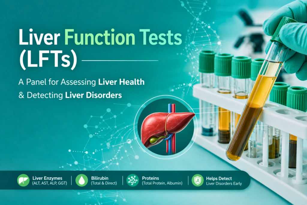
Table of Contents
Definition
Liver Function Tests (LFTs) are a battery of clinical biochemistry blood tests designed to evaluate the functional integrity of the liver, detect hepatic inflammation, and assess the patency of the biliary tract.
While called “function” tests, many components (like ALT and AST) actually measure cellular integrity rather than the liver’s synthetic or metabolic capacity.
Clinical Significance
LFTs are not merely “values on a report”; they are a multi-tool used to solve complex clinical puzzles. By analyzing the patterns of these markers, clinicians can pinpoint the exact nature of a liver pathology.
1. Detection of Hepatocellular Injury (The “Leaky Cell” Theory)
When hepatocytes are damaged by infection, toxins, or ischemia, their cell membranes become permeable, “leaking” enzymes into the bloodstream.
Viral Hepatitis: In acute viral hepatitis (A, B, or E), ALT and AST typically skyrocket, often exceeding 1,000 U/L.
Alcoholic Liver Disease: In these cases, we look for the De Ritis Ratio. Because alcohol depletes pyridoxal phosphate (a cofactor ALT needs), AST is often twice as high as ALT (AST:ALT > 2:1).
NAFLD/NASH: Non-Alcoholic Fatty Liver Disease usually presents with mild, chronic elevations where the ratio is closer to 1:1.
2. Assessment of Cholestasis (Biliary Roadblocks)
Cholestasis occurs when bile flow is interrupted. This triggers the synthesis of specific enzymes on the canalicular membrane of the liver cells.
Obstructive Jaundice: In cases like gallstones or pancreatic tumors, ALP and GGT rise disproportionately compared to ALT.
The GGT Confirmation: Since ALP can also come from bone, we use GGT as a “tie-breaker.” If ALP is high and GGT is also high, the source is definitely hepatic.
Bilirubin Partitioning: We look at Direct (Conjugated) Bilirubin. If it accounts for >50% of the total bilirubin, it strongly suggests a post-hepatic obstruction.
3. Monitoring Synthetic Function (The Factory Output)
This is the true measure of liver “function.” Unlike enzymes (which show damage), these markers show if the liver is actually doing its job.
Albumin (The Chronic Marker): With a half-life of 20 days, low albumin indicates chronic damage like Cirrhosis.
Prothrombin Time/INR (The Acute Marker): Clotting factors have short half-lives (hours). If the PT/INR is prolonged, it is an emergency signal of acute liver failure, as the “factory” has stopped producing essential proteins almost immediately.
4. Drug-Induced Liver Injury (DILI) Monitoring
Many common medications are hepatotoxic. We use the “R-Value” to categorize the injury:
Hepatocellular (R > 5): Seen with Acetaminophen or Isoniazid.
Cholestatic (R > 2): Seen with Anabolic steroids or Amoxicillin-clavulanate.
Hy’s Law: A critical clinical rule. If a patient has ALT > 3x ULN and Bilirubin > 2x ULN, there is a 10-50% mortality risk.
5. Staging Cirrhosis & Prognosis
In chronic liver disease, LFTs are fed into scoring systems to determine the patient’s survival rate or need for a transplant:
Child-Pugh Score: Uses Bilirubin, Albumin, and INR (along with clinical signs like ascites) to grade cirrhosis from Class A (well-compensated) to Class C (decompensated).
MELD Score (Model for End-Stage Liver Disease): A purely lab-based score using Bilirubin, INR, and Creatinine. This score is used globally to prioritize patients on liver transplant waiting lists.
Components of LFTs
The LFT panel is not a single test but a diagnostic mosaic. We categorize these markers based on whether they indicate leaking cells (injury), blocked pipes (cholestasis), or production failure (synthetic function).
1. Aminotransferases (The Hepatocellular Injury Markers)
These enzymes catalyze the interconversion of amino acids and ketoacids. When hepatocyte membranes are damaged, these enzymes leak into the systemic circulation.
Alanine Aminotransferase (ALT/SGPT):
Specificity: Highly specific to the liver.
Clinical Pearl: ALT has a longer half-life (~47 hours) than AST, making it a more persistent marker of acute injury.
Localization: Found primarily in the cytosol of hepatocytes.
Aspartate Aminotransferase (AST/SGOT):
The De Ritis Ratio (AST/ALT): * Ratio > 2:1: Strongly suggests Alcoholic Liver Disease (Alcohol suppresses ALT synthesis and damages mitochondria, releasing mitochondrial AST).
Ratio < 1: Typically seen in Viral Hepatitis or NAFLD.
Localization: Found in both mitochondria (80%) and cytosol (20%). It is also present in cardiac muscle, skeletal muscle, kidneys, and RBCs.
2. Alkaline Phosphatase (ALP) (The Cholestatic Marker)
ALP is a group of enzymes that hydrolyze phosphate esters at an alkaline pH.
Localization: Bound to the canalicular membrane of hepatocytes and the epithelium of bile ducts.
Elevation Mechanism: In biliary obstruction, bile salts accumulate and “detergent” the ALP off the membranes into the blood.
Non-Hepatic Sources: Bone (osteoblasts), placenta, and intestine. High ALP in a growing child or pregnant woman is often physiological, not pathological.
3. Gamma-Glutamyl Transferase (GGT) (The Confirmation Marker)
GGT is a microsomal enzyme involved in glutathione metabolism and the transport of amino acids across cell membranes.
Role in Differential Diagnosis: GGT is not found in bone. Therefore, if a patient has high ALP and high GGT, the source is hepatic. If GGT is normal, the ALP rise is likely from bone disease.
Induction: GGT is highly sensitive to enzyme-inducing substances, particularly alcohol and medications like phenytoin.
4. Bilirubin (The Excretory Marker)
Bilirubin is the end-product of heme degradation (mostly from old RBCs).
Unconjugated (Indirect) Bilirubin: Water-insoluble; bound to albumin.
High in: Hemolysis (overproduction) or Crigler-Najjar/Gilbert’s syndrome (impaired conjugation).
Conjugated (Direct) Bilirubin: Water-soluble; processed by the liver.
High in: Biliary obstruction (stones/tumors) or hepatocellular transport defects (Dubin-Johnson syndrome).
Delta Bilirubin: A fraction of conjugated bilirubin covalently bound to albumin; it explains why jaundice can persist long after an obstruction is cleared.
5. Markers of Synthetic Function (The True “Function” Tests)
While enzymes show damage, these markers measure the liver’s actual metabolic output.
Serum Albumin: The liver produces ~10-12g of albumin daily. Because its half-life is long (~20 days), it is a marker of chronic liver failure (Cirrhosis).
Prothrombin Time (PT/INR): The liver synthesizes clotting factors (I, II, V, VII, IX, X). Factor VII has a very short half-life (6 hours). Therefore, a prolonged PT is the earliest indicator of acute, severe liver damage (Fulminant Hepatic Failure).
Specimen Requirements
To ensure the integrity of the hepatic biomarkers, the following collection, handling, and stability protocols must be strictly followed.
1. Patient Preparation
Fasting Status: A 10–12 hour fast is strongly recommended.
Rationale: Post-prandial (after-meal) lipemia can cause significant turbidity, leading to interference in spectrophotometric assays (especially for ALT and Albumin).
Medication History: Patients should be advised to avoid hepatotoxic substances or enzyme-inducing drugs (like alcohol or certain anticonvulsants) for at least 24 hours prior to collection, as these can cause transient spikes in GGT.
2. Sample Collection (Tubes & Matrices)
The LFT panel is most commonly performed on Serum or Lithium Heparin Plasma.
| Tube Top Color | Additive | Matrix | Clinical Context |
| Gold / Red (SST) | Clot Activator & Gel | Serum | Most common for routine biochemistry. |
| Light Green (PST) | Lithium Heparin | Plasma | Preferred for “Stat” testing (no wait time for clotting). |
| Light Blue | Sodium Citrate | Plasma | Mandatory for Prothrombin Time (PT/INR). |
Critical Note: Avoid using EDTA (Purple top) or Oxalate tubes for LFTs. These anticoagulants chelate magnesium and zinc ions, which are essential cofactors for ALP activity, leading to falsely low results.
3. Handling and Specimen Integrity (The “Big Three” Interferences)
As a technologist, you must visually inspect every sample for the following:
Hemolysis (Red Serum): Reject samples with significant hemolysis.
Effect: RBCs contain 40x more AST and 7x more ALT than serum. Rupture of these cells will cause a massive false elevation.
Icterus (Dark Yellow/Orange Serum): High bilirubin levels (icterus) can cause spectral interference in assays that measure absorbance at wavelengths near 450-500 nm.
Lipemia (Milky Serum): Caused by triglycerides. Lipemia scatters light in the analyzer’s flow cell, falsely increasing absorbance readings for Total Protein and Albumin.
4. Storage and Stability
Liver enzymes and pigments are sensitive to environmental conditions.
Bilirubin (Light Sensitivity): Bilirubin is highly photo-labile. Exposure to sunlight or fluorescent lab lights can decrease bilirubin concentration by up to 50% per hour.
Action: Store samples in amber tubes or wrap them in aluminum foil.
Temperature Stability:
Room Temp (18–25°C): Stable for 1–2 days.
Refrigerated (2–8°C): Stable for 7 days (Preferred).
Frozen (-20°C): Stable for months (Required for long-term research).
ALP Recovery: Interestingly, ALP activity can increase slightly (2-3%) after refrigeration or standing, as the enzyme slowly reactivates.
5. Centrifugation
Timing: Serum should be separated from the clot within 2 hours of collection.
Speed: Typically 3000-3500 RPM for 10 minutes.
Rationale: Prolonged contact with RBCs can lead to the leakage of intracellular enzymes (LDH, AST) and shifts in pH that affect enzyme stability.
Summary Checklist for the Lab Bench
Verify Patient ID: Match name/DOB on tube and requisition.
Check Tube Type: Ensure SST or Lithium Heparin was used.
Screen for Hemolysis: Reject if the serum is visibly pink or red.
Protect from Light: Ensure bilirubin samples are covered.
Centrifuge Properly: Ensure a clear separation of serum/plasma from cells.
Methodology & Principal
Modern clinical chemistry analyzers utilize standardized enzymatic and colorimetric assays. As a technologist, understanding these reactions allows you to identify potential interferences and validate the accuracy of the results.
1. Aminotransferases (ALT & AST)
Methodology: Kinetic UV (Standardized by IFCC).
Principle: These assays use a coupled enzymatic reaction.
For ALT: L-Alanine + alpha-ketoglutarate —ALT—> Pyruvate + L-Glutamate. The pyruvate is then reduced to Lactate by Lactate Dehydrogenase (LDH), while NADH is oxidized to NAD+.
For AST: L-Aspartate + alpha-ketoglutarate —AST—> Oxaloacetate + L-Glutamate. The oxaloacetate is reduced to Malate by Malate Dehydrogenase (MDH), with NADH oxidation.
Measurement: The rate of decrease in absorbance is measured at 340 nm as NADH is consumed. The rate of decrease is directly proportional to the enzyme activity in the sample.
2. Alkaline Phosphatase (ALP)
Methodology: Kinetic Colorimetric (Bowers and McComb).
Principle: ALP catalyzes the hydrolysis of p-nitrophenyl phosphate (pNPP) in an alkaline environment (pH 10.4) in the presence of magnesium and zinc ions.
Reaction: p-Nitrophenyl phosphate + H2O —ALP—> p-Nitrophenol (Yellow) + Phosphate.
Measurement: The rate of formation of p-Nitrophenol (a yellow-colored compound) is measured spectrophotometrically at 405 nm.
3. Bilirubin (Total & Direct)
Methodology: Jendrassik-Grof Method (The Gold Standard).
Principle: Bilirubin reacts with diazotized sulfanilic acid to form a purple-colored compound called azobilirubin.
Direct (Conjugated): Reacts rapidly with the diazo reagent in an aqueous medium.
Total: Requires an “accelerator” (solubilizer) like Caffeine-benzoate or Methanol to release the unconjugated bilirubin from albumin so it can react with the diazo reagent.
Reaction Termination: Ascorbic acid is added to stop the reaction, and an alkaline tartrate is added to turn the purple azobilirubin into a blue-green color, which is then measured at 600 nm.
4. Albumin
Methodology: Dye-Binding Method (Bromocresol Green – BCG).
Principle: In a buffered acidic environment (pH 4.2), Albumin acts as a cation and binds with the anionic dye Bromocresol Green.
Color Change: The binding causes a shift in the absorption maximum of the dye, changing it from yellow-green to blue-green.
Measurement: The intensity of the blue-green color is measured at 628 nm and is directly proportional to the albumin concentration.
5. Total Protein
Methodology: Biuret Method.
Principle: Cupric ions (Cu2+) in an alkaline solution react with the peptide bonds of proteins (at least two peptide bonds are required).
Complex Formation: This creates a violet-colored chelate complex.
Measurement: The absorbance of the violet complex is measured at 540 nm. The intensity is proportional to the number of peptide bonds present, and thus, the protein concentration.
Technologist’s Summary Table
| Analyte | Wavelength | Key Reagent/Enzyme | Potential Interference |
| ALT/AST | 340 nm | NADH | Hemolysis (Falsely increases AST) |
| ALP | 405 nm | pNPP | Hemolysis (Inhibition) |
| Bilirubin | 600 nm | Diazo / Caffeine | Light (Photodegradation) |
| Albumin | 628 nm | Bromocresol Green | Lipemia (Turbidity) |
| Protein | 540 nm | Copper Sulfate | Lipemia / Hemolysis |
Normal Ranges & Interpretations
Note: Reference ranges may vary slightly depending on the laboratory’s equipment and population.
| Test | Normal Range (Adult) | Increased Levels Indicate | Clinical Interpretation |
| ALT (SGPT) | 7–55 U/L | Viral hepatitis, fatty liver, toxins | Specific marker for liver injury |
| AST (SGOT) | 8–48 U/L | Cirrhosis, muscle trauma, MI | Non-specific; check AST/ALT ratio |
| ALP | 40–129 U/L | Cholestasis, Paget’s disease | Marker of biliary/bone activity |
| Bilirubin (Total) | 0.1–1.2 mg/dL | Jaundice, hemolysis, obstruction | Excretory function marker |
| Albumin | 3.5–5.0 g/dL | Dehydration | Reflects long-term synthetic capacity |
| Prothrombin Time | 11–13.5 sec | Acute/Chronic liver failure | Acute marker of synthetic function |
Quick Stats
| Feature | Details | Things You Need to Know |
| Test Type | Clinical Biochemistry | LFTs are a diagnostic “mosaic” assessing three areas: Hepatocellular injury (Enzymes), Cholestasis (Bilirubin/ALP), and Synthetic function (Albumin/PT). |
| Sample Type | Routine Venous Draw | Collected in a Gold Top (SST) or Lithium Heparin (Green) tube. Plasma is preferred for “Stat” emergency orders. |
| Fasting Required? | Strongly Recommended | A 10–12 hour fast prevents lipemia, which can interfere with spectrophotometric readings of Albumin and Total Protein. |
| Sample Integrity | Light Protection | Bilirubin is photo-labile. Samples must be wrapped in foil or stored in amber tubes to prevent a false decrease in values (up to 50% loss per hour). |
| Turnaround Time | 30–60 Minutes | Analyzed via automated kinetic UV and colorimetric assays. Critical for diagnosing acute liver failure or toxic ingestion (e.g., Acetaminophen). |
| Primary Metric | De Ritis Ratio (AST/ALT) | This calculated ratio helps distinguish between alcoholic liver disease (>2.0) and viral/non-alcoholic fatty liver disease (<1.0). |
| Clinical Purpose | Screening & Differential | Used to screen for hepatitis, monitor hepatotoxic drugs (Statins), and stage cirrhosis using the Child-Pugh or MELD scores. |
| Critical Values | ALT/AST >1000 U/L | Levels this high are “panic values” indicating acute viral hepatitis, ischemic shock liver, or severe toxic injury. |
Strategic Considerations
The accuracy of an LFT report and its subsequent clinical action depends on a seamless flow between the collector, the analyzer, and the prescriber.
1. Considerations for Lab Technicians (The Gatekeepers)
The technician is responsible for the Pre-Analytical and Analytical phases. Your focus is on sample integrity.
Visual Inspection: Always check for Hemolysis, Icterus, and Lipemia (HIL). If a sample is moderately hemolysed, request a redraw, as the AST result will be clinically invalid.
Timely Centrifugation: Ensure serum is separated from cells within 2 hours to prevent leakage of intracellular enzymes.
Light Protection: Bilirubin is your most fragile analyte. Keep the sample shielded from ambient lab light to prevent photodegradation.
Instrument Maintenance: Perform daily “Start-up” and Quality Control (QC) for the chemistry analyzer. If QC for ALT or ALP is out of range ($2SD$), do not process patient samples until the issue is resolved.
2. Considerations for Lab Technologists (The Quality Experts)
The technologist oversees the Analytical validation and Post-Analytical troubleshooting.
Result Correlation: Practice “Delta Checks.” If a patient’s ALT was 40 U/L yesterday and is 400 U/L today, investigate—is it a real clinical spike (e.g., acute drug toxicity) or a mislabeled sample?
Linearity & Dilutions: If an ALT result exceeds the analyzer’s linearity (e.g., >2000 U/L), perform a manual 1:10 dilution with saline and multiply the result accordingly.
Interference Management: If the sample is lipemic, use a high-speed centrifuge (ultracentrifuge) to clear the lipids or use a methodology (like an enzymatic albumin assay) less prone to turbidity interference.
Critical Value Reporting: Immediately notify the physician if enzymes exceed panic levels (e.g., >1000 U/L) or if the Bilirubin is critically high in a neonate (Risk of Kernicterus).
3. Considerations for Doctors (The Clinical Interpreters)
The physician focuses on the Diagnostic Logic and patient management.
Look for Patterns, Not Single Numbers: An isolated mild rise in AST is rarely liver disease; it may be skeletal muscle injury from intense exercise. Always look for the Pattern (Hepatocellular vs. Cholestatic).
Contextualize with Patient History: Before diagnosing Hepatitis, check for recent medication changes (Statins, NSAIDs) or herbal supplements that might cause Drug-Induced Liver Injury (DILI).
Assess Function vs. Injury: Remember that ALT/AST tell you about cell death, but Albumin and INR tell you if the patient is in liver failure. A patient can have normal enzymes but still be in end-stage cirrhosis.
Standardized Scoring: For chronic patients, use the LFT data to calculate MELD or Child-Pugh scores to determine the urgency of specialist referral or transplant.
FAQs
1. What are the 7 tests in a liver function panel?
A standard Liver Function Test (LFT) panel typically includes:
ALT (Alanine Aminotransferase)
AST (Aspartate Aminotransferase)
ALP (Alkaline Phosphatase)
GGT (Gamma-Glutamyl Transferase)
Total Bilirubin
Albumin
Total Protein Note: Some labs also include Prothrombin Time (PT/INR) to assess acute synthetic function.
2. What does it mean if my ALT is higher than my AST?
When ALT is higher than AST, it usually indicates a hepatocellular pattern of injury. This is commonly seen in Viral Hepatitis, Non-Alcoholic Fatty Liver Disease (NAFLD), or exposure to certain toxins. Since ALT is more liver-specific, its elevation is a primary indicator of hepatic inflammation.
3. Can you have liver disease with normal LFT results?
Yes. Chronic liver conditions, such as compensated cirrhosis or fatty liver, can sometimes show normal enzyme levels because the liver has stopped “leaking” enzymes or has significant scarring. This is why clinical correlation with imaging (Ultrasound/FibroScan) is essential.
4. Why is GGT tested along with ALP?
GGT is used to determine the origin of an elevated ALP. If both GGT and ALP are high, the problem is likely in the liver or bile ducts. If ALP is high but GGT is normal, the elevation is likely coming from bone disease, pregnancy, or the intestines.
5. Does a high bilirubin always mean liver failure?
Not necessarily. High unconjugated bilirubin can be caused by hemolysis (excessive breakdown of red blood cells) or Gilbert’s Syndrome (a benign genetic condition). High conjugated bilirubin, however, more often points to bile duct obstruction or significant liver damage.
6. How does alcohol affect LFT results?
Alcohol consumption typically causes a “flipped” ratio where AST is at least twice as high as ALT (AST:ALT > 2:1). It also significantly induces the production of GGT, making it a sensitive marker for heavy alcohol use.
7. Why do I need to fast before a Liver Function Test?
Fasting (usually 8–12 hours) is preferred to prevent lipemia (excess fat in the blood). Lipemia makes the serum cloudy, which can interfere with the optical sensors in lab analyzers, leading to inaccurate readings for Albumin and Total Protein.