
Clotting Time
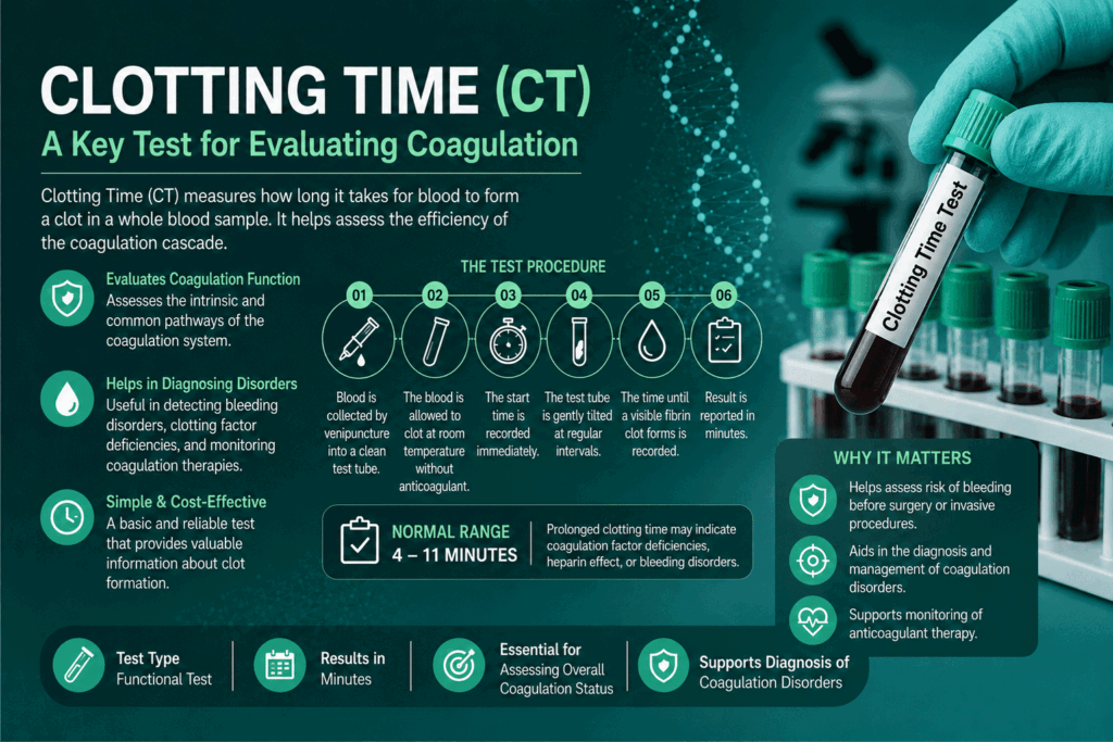
Table of Contents
Definition
Clotting time (CT) is a traditional screening test that measures the period required for a sample of whole blood to coagulate (form a solid fibrin clot) under strictly controlled in vitro conditions. Unlike plasma-based automated tests, traditional CT assays evaluate clotting in its native state, relying entirely on the interaction of the patient’s own cellular elements, platelets, and plasma coagulation factors.
Clinical Significance
Here is an advanced, comprehensive breakdown of the clinical significance of clotting time, organized by physiological pathway, specific factor thresholds, and complex clinical states.
1. What CT Tells the Lab
Clotting time specifically measures the functionality of the intrinsic and common coagulation pathways. It triggers via contact activation when the plasma proteins interact with a negatively charged surface (like glass).
If a patient exhibits a prolonged CT, the underlying pathology must be systematically mapped back to this cascade:
Intrinsic Pathway Factors: Factor XII (Hageman factor), Factor XI (Plasma thromboplastin antecedent), Factor IX (Christmas factor), and Factor VIII (Antihemophilic factor).
Common Pathway Factors: Factor X (Stuart-Prower factor), Factor V (Proaccelerin), Factor II (Prothrombin), and Factor I (Fibrinogen).
2. Pathological Breakdown of Prolonged Clotting Time
A. Congenital Hemoglobinopathies and Factor Deficiencies
The most common genetic reasons for a prolonged CT are structural or quantitative deficiencies in specific intrinsic factors:
Hemophilia A (Classic Hemophilia): An X-linked recessive disorder resulting in a deficiency of Factor VIII.
Hemophilia B (Christmas Disease): An X-linked recessive disorder causing a deficiency of Factor IX.
Hemophilia C: An autosomal recessive deficiency of Factor XI, seen predominantly in Ashkenazi Jewish populations.
Factor XII Deficiency: Interestingly, a deficiency in Factor XII (or its cofactors Prekallikrein and High-Molecular-Weight Kininogen) markedly prolongs clotting time in vitro, but does not cause clinical bleeding in vivo. This is a crucial distinction for board exams.
The Sensitivity Threshold Warning: Manual clotting time is a relatively insensitive assay. A patient’s factor levels must typically drop below 1% to 5% of normal activity for the Lee-White or capillary methods to register as prolonged. Mild or moderate hemophilia will frequently return a completely normal clotting time.
B. Acquired Pathologies and Organ Dysfunction
Severe Hepatic Failure: The parenchyma of the liver synthesized nearly all coagulation factors (except for von Willebrand Factor and parts of Factor VIII). Advanced cirrhosis or acute liver necrosis disrupts this synthesis, causing a steep decline in common pathway factors (Prothrombin, Factor V, Factor X) and a consequently prolonged CT.
Vitamin K Deficiency: Factors II, VII, IX, and X require Vitamin K for gamma-carboxylation, allowing them to bind to phospholipid surfaces. A deficiency due to malabsorption, prolonged antibiotic therapy, or dietary lack cripples both the intrinsic (IX) and common (II, X) pathways.
C. Therapeutic and Accidental Anticoagulation
Unfractionated Heparin (UFH) Therapy: Heparin binds to Antithrombin III, accelerating its ability to inactivate Thrombin (IIa) and Factor Xa by over 1,000-fold. This immediate, profound arrest of the common pathway intensely prolongs clotting time.
Warfarin (Coumadin) Toxicity: As a Vitamin K antagonist, severe Warfarin overdoses deplete the active forms of factors II, IX, and X, leading to prolonged CT.
D. Consumption Coagulopathies
Disseminated Intravascular Coagulation (DIC): Triggered by severe trauma, sepsis, or malignancy, DIC causes systemic, uncontrolled microvascular clotting. This massive, runaway process rapidly consumes the body’s circulating pool of Fibrinogen (Factor I), Prothrombin (Factor II), and platelets, leaving the blood depleted of the basic elements required to clot. The resulting state shows severely prolonged clotting times alongside catastrophic systemic bleeding.
3. Why Clotting Time Remains Normal in Platelet & Extrinsic Disorders
Understanding what the test cannot detect is just as clinically valuable as knowing what it can.
Platelet Deficiencies (Thrombocytopenia or Glanzmann Thrombasthenia): While platelets provide the phospholipid scaffolding for optimal in vivo clotting, manual whole-blood clotting tests have enough background tissue/surface activation to bypass mild platelet issues. The fibrin mesh will still form on time, meaning CT is entirely normal in platelet disorders.
Extrinsic Pathway Deficiencies (Factor VII): Factor VII operates purely in the extrinsic pathway (activated by Tissue Factor). Because the glass tube assay relies purely on contact (intrinsic) activation, an isolated Factor VII deficiency will never prolong a clotting time test.
4. Clinical Significance of Shorter-Than-Normal Clotting Time
A hypercoagulable state where blood clots faster than the standard reference ranges (e.g., less than 5 minutes in the Lee-White method) is less common but highly notable:
Disseminated Malignancy: Certain adenocarcinomas secrete procoagulant mucins or tissue-factor-like substances directly into circulation, inducing a systemic hypercoagulable state.
Acute Thrombophlebitis / Early DIC: The hypercoagulable “thrombotic phase” immediately preceding the consumption phase of DIC can exhibit accelerated clot formation.
Pre-analytical Errors: Most short clotting times are artifacts caused by poor draw technique, such as a traumatic venipuncture introducing external tissue thromboplastin into the needle barrel.
5. Diagnostic Interpretation Matrix
When evaluating an abnormal clotting time in conjunction with modern screening assays, clinicians look at the complete profile:
| Clotting Time (CT) | Bleeding Time (BT) | PT (Extrinsic) | aPTT (Intrinsic) | Suspected Clinical Condition |
| Prolonged | Normal | Normal | Prolonged | Hemophilia A or B; Heparin therapy |
| Prolonged | Prolonged | Prolonged | Prolonged | Severe Liver Disease; Consumptive DIC |
| Normal | Prolonged | Normal | Normal | Severe Thrombocytopenia; Von Willebrand Disease |
| Normal | Normal | Prolonged | Normal | Early Vitamin K Deficiency; Isolated Factor VII deficiency |
Methods and Principle
Understanding the detailed scientific principles and execution protocols for clotting time assays guarantees accuracy in manual hematology. If your procedure, materials, or environment shift even slightly, your manual results will skew.
The Fundamental Scientific Principle of Clotting Time
The underlying principle of the clotting time test is surface-induced contact activation of the intrinsic coagulation pathway.
When fresh, whole, unanticoagulated blood is removed from the vascular system and exposed to a foreign, non-endothelial surface (such as the glass wall of a test tube or capillary tube), the cascade begins without any added chemical triggers.
[Negatively Charged Glass Surface]
              │
              ▼
    Factor XII  ──>  Factor XIIa (Contact Activation)
              │
              ▼
    Factor XI   ──>  Factor XIa
              │
              ▼
    Factor IX   ──>  Factor IXa (Combines with VIIIa)
              │
              ▼
    Factor X    ──>  Factor Xa  (Enters Common Pathway)
              │
              ▼
    Prothrombin (II) ──> Thrombin (IIa)
              │
              ▼
    Fibrinogen  (I)  ──> Fibrin Gel Clot
Contact Activation: The negatively charged silanol groups present on raw glass surfaces act as a catalyst. They induce a conformational change in Factor XII (Hageman Factor), converting it into its active enzymatic form, Factor XIIa.
Cascade Progression: Factor XIIa activates Factor XI, which subsequently activates Factor IX. Factor IXa forms a complex with Factor VIIIa, calcium ions, and platelet phospholipids to activate Factor X, entering the common pathway.
Thrombin Generation: The prothrombinase complex (Factors Xa, Va, and calcium) converts Prothrombin (Factor II) into Thrombin (Factor IIa).
Fibrin Polymerization: Thrombin cleaves soluble Fibrinogen (Factor I) into insoluble Fibrin monomers. These monomers polymerize into a dense, cross-linked meshwork that traps erythrocytes and platelets, transforming the liquid blood into a solid gel clot.
Because no chemical reagents, tissue factor, or anticoagulants are added, the time it takes for this solid clot to form is entirely dependent on the concentration and functional integrity of the patient’s own intrinsic and common pathway factors.
Method 1: The Lee-White Method (Macro-Tube Technique)
The Lee-White method is the classic standard for manual clotting time because it uses venous blood, limiting the interference of tissue juices.
Reagents and Equipment
Water bath calibrated and maintained strictly at 37°C ± 0.5°C.
Three clean, dry, plain glass test tubes (standard 10 × 75 mm or 13 × 100 mm). Plastic tubes cannot be used.
Sterile syringe, 21-gauge needle, and venipuncture supplies.
Stopwatch.
Standard Operating Procedure (SOP)
1.Pre-Warming:Reduces temperature variables.
Place the three labeled glass test tubes (Tube 1, Tube 2, and Tube 3) into the 37°C water bath. Coagulation factors are highly temperature-sensitive; cold tubes falsely prolong results.
2.Clean Venipuncture:Crucial for timing accuracy.
Perform a clean, swift venipuncture. Start the stopwatch the exact moment blood begins entering the syringe. A traumatic draw forces tissue thromboplastin into the needle, activating the extrinsic pathway and causing a falsely shortened clotting time.
3.Sample Distribution:1.0 mL aliquots.
Remove the needle from the syringe and gently dispense exactly 1.0 mL of whole blood down the inner wall of each pre-warmed tube. Gentle dispensing prevents mechanical hemolysis and premature platelet activation. Place them immediately back in the water bath.
4.Sequential Tilting:Begins at Minute 3.
Leave the tubes completely undisturbed for the first 3 minutes. At exactly 3 minutes, remove Tube 1 from the water bath and tilt it gently to a 45° angle. If blood still flows, return it to the water bath. Repeat this gentle tilt on Tube 1 every 30 seconds.
5.Progressive Assessment:Prevents mechanical disruption.
As soon as Tube 1 clots completely (solidifies so that it can be inverted without spilling), record the time. Immediately begin the same 30-second tilting process with Tube 2. Once Tube 2 clots, immediately move to Tube 3.
6.Endpoint Determination:The official test result.
The official clotting time is recorded the exact second the blood in Tube 3 forms a complete, solid gel clot.
Normal Reference Range: 5 to 15 minutes.
Reporting: Results are reported rounded to the nearest half-minute.
Method 2: The Capillary Tube Method (Dale and Laidlaw Technique)
This technique is primarily used in pediatric or field settings where venipuncture is difficult or resources are limited. It carries a higher margin of error due to unavoidable tissue factor contamination.
Reagents and Equipment
Non-heparinized, plain glass capillary tubes (blue tip, roughly 75 mm long, 1 mm internal diameter).
Sterile lancet and alcohol prep pads.
Stopwatch.
Standard Operating Procedure (SOP)
Fingertip Puncture: Clean the patient’s finger (or heel for infants) with alcohol. Allow it to air-dry completely. Puncture the skin deeply enough with the lancet to ensure a free, spontaneous flow of blood.
Start Timing: Wipe away the very first drop of blood with dry gauze (it contains highly concentrated tissue fluid). Start the stopwatch the moment the second drop of blood forms.
Fill the Tube: Hold the non-heparinized capillary tube horizontally and bring it into contact with the blood drop. Allow the tube to fill to at least 74% of its length via capillary action.
Incubation: Hold the tube between your palms or place it inside a portable incubator to keep it near body temperature (~37°C).
Sequential Snapping: Starting at exactly 2 minutes, carefully snap a small 5 mm piece off the end of the capillary tube. Pull the broken pieces apart slowly (about 1–2 cm away from each other).
Fibrin Strand Endpoint: Repeat this snapping process every 30 seconds. The test is complete the exact moment a visible, thread-like fibrin strand spans the gap between the two broken ends of the glass tube.
Normal Reference Range: 2 to 6 minutes.
Technical Troubleshooting Matrix
To avoid generating erratic manual data, you must carefully monitor the technical variables highlighted below:
| Technical Variable | Pathophysiological Impact | Corrective Laboratory Action |
| Mechanical Agitation | Continual tilting breaks early, fragile fibrin strands, resetting the network formation and falsely prolonging the clotting time. | Leave Tubes 2 and 3 completely stationary while checking Tube 1. |
| Surface-Area-to-Volume Ratio | Wider tubes decrease the glass-to-blood ratio, while narrow tubes increase it. Higher relative surface contact artificially accelerates Factor XII activation. | Standardize tube dimensions exactly (typically 10 × 75 mm) to match validated reference ranges. |
| Plastic Material Use | Plastic tubes lack the negative charge required to alter Factor XII conformation, preventing contact activation. | Ensure the use of plain, additive-free glass tubes exclusively. |
| Temperature Deviations | Coagulation is an enzymatic process. Dropping below 35°C significantly delays enzyme kinetics, leading to a falsely prolonged result. | Regularly check water bath calibration during testing. |
Specimen Requirements
For automated coagulation testing, specimen collection typically involves drawing blood into a sodium citrate tube, followed by centrifugation to isolate platelet-poor plasma. However, manual clotting time (CT) assays follow a completely unique set of rules: the specimen is tested in its raw, unanticoagulated, whole-blood state immediately upon collection.
Because there is no preservative or anticoagulant to halt chemical degradation, the margin for pre-analytical error is remarkably slim. Below is the comprehensive breakdown of specimen requirements and collection protocols for clotting time tests.
1. Type of Specimen required
| Method | Required Specimen Matrix | Collection Volume |
| Lee-White Method | Fresh, non-anticoagulated venous whole blood | Exactly 3.0 mL (divided evenly into three 1.0 mL aliquots) |
| Capillary Tube Method | Fresh, non-anticoagulated capillary whole blood | ~70–100 microliters |
Critical Rule: The specimen cannot be stored, refrigerated, or batched. The time window between drawing the blood from the patient’s vessel and initiating the test is zero seconds. The test begins the moment the needle enters the lumen of the vein.
2. Patient Preparation and Screening Prerequisite
Before performing the collection, the phlebotomist or laboratory scientist must verify the patient’s status to ensure specimen integrity:
Fasting Status: While fasting is not strictly mandatory for a coagulation assay, a heavily lipemic specimen (caused by a recent high-fat meal) can obscure the optical assessment of early fibrin strands, especially in micro-capillary testing.
Anticoagulant Review: It is vital to document if the patient is currently receiving Unfractionated Heparin (UFH), Warfarin (Coumadin), or Direct Oral Anticoagulants (DOACs). If the draw is meant to establish a baseline or clear a patient for surgery, these medications will drastically skew the specimen’s performance.
Resting State: The patient should be at rest for at least 15 minutes before the procedure. Severe physical exertion or extreme emotional stress triggers the release of endogenous epinephrine, which transiently elevates Factor VIII and Von Willebrand Factor levels, potentially shortening the baseline clotting time.
3. Venous Blood Collection Protocol (Lee-White Specific)
The venipuncture protocol for manual clotting time is highly specific. Any variation can result in a compromised specimen that must be rejected.
Syringe vs. Evacuated Tube System (ETS)
For the classic manual Lee-White method, a sterile syringe system is heavily preferred over a standard vacuum tube system. The sudden, high-pressure vacuum shear force inside an evacuated tube can prematurely rupture platelets, releasing platelet factor 3 (PF3) into the specimen and artificially accelerating the clotting cascade.
Needle Gauge Selection
Standard Selection: A 21-gauge or 22-gauge needle must be used.
Why it matters: A needle that is too narrow (e.g., 25-gauge) creates high shear stress, physically shearing red blood cells (hemolysis) and prematurely activating clotting factors. A needle that is too wide can cause excessive localized tissue trauma, flooding the early blood flow with tissue thromboplastin.
Order of Draw and Tissue Thromboplastin Management
When a needle passes through human skin and subcutaneous tissue, it inevitably cuts through cells, collecting a small amount of tissue thromboplastin (Tissue Factor) inside the tip of the needle. Tissue factor activates the extrinsic pathway (Factor VII), which completely bypasses the intrinsic pathway you are trying to measure.
[Skin Puncture] ──> Extrinsic Tissue Factor enters needle ──> Bypasses Intrinsic Assay ──> Falsely Shortened CT
To eliminate this pre-analytical contamination:
If using a syringe exclusively for this test, some advanced clinical protocols recommend performing a two-syringe technique. Draw the first 1–2 mL of blood into a primary syringe (and discard it or use it for other tests), then swap syringes while leaving the needle hub in place to draw the pristine 3 mL clotting time specimen.
If drawing as part of a multi-tube panel using an Evacuated Tube System, the clotting time sample cannot be the first tube drawn. You must draw a waste tube or a routine biochemistry/hematology tube first to clear the tissue factor out of the needle lumen.
4. Capillary Blood Collection Protocol (Micro-Method Specific)
If executing the capillary tube method, the requirements focus entirely on establishing a free-flowing vascular bed:
Site Selection: The palmar surface of the distal phalanx of the ring or middle finger is preferred for adults; the lateral or medial plantar surface of the heel is mandatory for infants.
Temperature Optimization: The collection site must be warm. If the hand is cold, capillaries constrict, causing poor blood flow. Warm the site for 3–5 minutes using a warm compress to ensure a brisk, spontaneous flow.
Wiping the First Drop: The very first drop of capillary blood following a skin puncture contains highly concentrated tissue fluid from the disrupted dermis. This first drop must be wiped away with clean, dry gauze. Only the subsequent, freely flowing drops are acceptable for collection into the plain glass capillary tube.
Prohibition of “Milking”: You must never excessively squeeze, massage, or “milk” the puncture site to force blood out. Squeezing forces interstitial fluid and intracellular contents out of the surrounding tissues, diluting the plasma proteins and introducing tissue factor, which invalidates the specimen.
5. Specimen Rejection Criteria
In a clinical laboratory setting, any specimen exhibiting the following characteristics must be discarded, and a re-draw must be requested immediately:
Presence of Clots Prior to Testing: If visible micro-clots or fibrin strings are observed in the syringe or capillary tube before timing begins, the coagulation cascade has already progressed past its initial stages.
Hemolysis: Hemolyzed samples release intracellular erythrocyte contents (such as adenosine diphosphate and red cell stroma) that act as thromboplastin-like substances, invalidates the assay.
Anticoagulant Contamination: The unintentional use of a EDTA (lavender), Sodium Citrate (light blue), or Heparin (green) collection device. The specimen must be completely additive-free.
Inappropriate Temperature Maintenance: Any specimen allowed to sit at room temperature or exposed to drafts prior to testing, rather than being incubated in a 37°C environment.
Reference Ranges and Clinical Interpretations
1. Reference Ranges for Clotting Time
Reference values for manual clotting time assays are heavily dependent on the methodology, equipment dimensions, and temperature control used. Because these tests evaluate whole blood in its native state, the surface-area-to-volume ratio dictates the normal boundaries.
| Method | Normal Reference Range | Temperature Standard | Specimen Source |
| Lee-White Method (Tube) | 5 to 15 minutes | Strictly 37°C | Venous Whole Blood |
| Capillary Tube Method | 2 to 6 minutes | Body Temperature (~37°C) | Capillary Whole Blood |
Critical Laboratory Note: There is no universal, standardized reference range for manual assays. Every clinical laboratory must establish and validate its own internal reference intervals based on its specific tube dimensions, protocol, and technician baseline.
2. Clinical Interpretation Matrix
When interpreting clotting time results, a prolonged or shortened interval must be systematically correlated with specific pathway failures or pre-analytical errors.
                  ┌──────────────────────┐
                  │ Clotting Time Result │
                  └──────────┬───────────┘
                             │
            ┌────────────────┴────────────────┐
            ▼                                 ▼
   [Prolonged (>15 min)]             [Shortened (<5 min)]
            │                                 │
     Factor Deficiencies               Pre-Analytical Errors
     (VIII, IX, XI, XII)               (Traumatic draw, Tissue factor)
            │                                 │
     Circulating Inhibitors            Hypercoagulability
     (Heparin, Lupus Anticoagulant)     (Disseminated Malignancy)
A. Prolonged Clotting Time (>15 minutes in Lee-White)
A prolonged clotting time indicates that the enzymatic cascade responsible for generating a solid fibrin mesh is compromised. This occurs due to severe factor deficiencies, active drug therapy, or consumption states.
1. Severe Congenital Coagulation Factor Deficiencies
Factor VIII Deficiency (Hemophilia A): Causes a marked delay in the intrinsic tenase complex (Factor VIIIa + Factor IXa + Calcium + Phospholipids), hindering the activation of Factor X.
Factor IX Deficiency (Hemophilia B): Directly halts the intrinsic pathway due to a lack of the functional protease needed to activate Factor X.
Factor XI Deficiency (Hemophilia C): Results in delayed activation of Factor IX, leading to slower downstream thrombin generation.
Factor XII Deficiency (Hageman Factor): Causes profound prolongation in vitro because the glass surface cannot initiate the contact activation phase. Clinical note: This does not translate to bleeding risks in the patient.
Common Pathway Deficiencies (Factors X, V, II, or I): Severe deficiencies in Prothrombin (II) or Fibrinogen (I) mean that even if the intrinsic pathway operates perfectly, the machinery to build the actual physical clot structure is missing.
The Sensitivity Threshold Warning: Manual clotting time is a relatively insensitive assay. A patient’s factor levels must typically drop below 1% to 5% of normal activity for the Lee-White or capillary methods to register as prolonged. Mild or moderate hemophilia will frequently return a completely normal clotting time.
2. Therapeutic Anticoagulation and Inhibitors
Unfractionated Heparin (UFH): Heparin binds to Antithrombin III, causing immediate inactivation of Thrombin (IIa) and Factor Xa. This completely stalls the common pathway, frequently pushing clotting times well past 30 minutes.
Circulating Anticoagulants/Inhibitors: Specific factor inhibitors (e.g., anti-Factor VIII antibodies) or non-specific inhibitors like the Lupus Anticoagulant bind to and neutralize clotting components.
3. Acquired Systemic Pathologies
Advanced Liver Disease: The liver parenchymal cells synthesize nearly all clotting factors. Cirrhosis, hepatitis, or acute liver failure leaves the body with an insufficient concentration of Factors II, V, IX, and X to execute clotting within standard intervals.
Severe Vitamin K Deficiency: Bypasses the synthesis of functional factors II, IX, and X, altering both intrinsic and common pathway kinetics.
Consumptive Coagulopathy (DIC): Systemic microvascular clotting consumes the body’s circulating pool of Fibrinogen (Factor I), Prothrombin (Factor II), and platelets, leaving the blood depleted of the basic elements required to clot.
B. Shortened Clotting Time (<5 minutes in Lee-White)
A hypercoagulable state where blood clots faster than the standard reference ranges is less common but highly notable:
Pre-analytical Errors: Most short clotting times are artifacts caused by poor draw technique, such as a traumatic venipuncture introducing external tissue thromboplastin into the needle barrel.
Disseminated Malignancy: Certain adenocarcinomas secrete procoagulant mucins or tissue-factor-like substances directly into circulation, inducing a systemic hypercoagulable state.
Acute Thrombophlebitis / Early DIC: The hypercoagulable “thrombotic phase” immediately preceding the consumption phase of DIC can exhibit accelerated clot formation.
3. Diagnostic Interpretation Profile
To help differentiate between overlapping bleeding disorders, clinical pathology relies on cross-referencing multiple panels:
| Condition | Clotting Time (CT) | Bleeding Time (BT) | Prothrombin Time (PT) | Activated Partial Thromboplastin Time (aPTT) |
| Severe Hemophilia A/B | Prolonged | Normal | Normal | Prolonged |
| Severe Liver Failure / Advanced DIC | Prolonged | Prolonged | Prolonged | Prolonged |
| Thrombocytopenia | Normal | Prolonged | Normal | Normal |
| Isolated Factor VII Deficiency | Normal | Normal | Prolonged | Normal |
| Unfractionated Heparin Therapy | Prolonged | Normal | Normal or Prolonged | Severe Prolongation |
Quick Stats
| Feature | Details | Critical Lab Insights |
| Test Type | Coagulation / Functional Secondary Hemostasis Marker | A direct in vitro indicator of the intrinsic and common coagulation pathways. It evaluates the blood’s native ability to generate a stable fibrin mesh without added chemical activators. |
| Sample Type | Fresh, Native whole blood (Unanticoagulated) | Tested immediately upon collection without preservatives, anticoagulants, or centrifugation. The sample is completely non-storable and must be analyzed in real-time. |
| Method Standard | Lee-White (Macro-Tube) Method | The manual benchmark. It utilizes three pre-warmed glass tubes maintained at 37°C to evaluate contact activation, minimizing tissue thromboplastin interference compared to capillary methods. |
| Primary Metric | Elapsed Time (Minutes) | Measured from the exact second venous blood enters the syringe barrel to the complete solidification of blood in the third test tube. |
| Patient Prep Window | Active Anticoagulant Assessment | Current heparin, warfarin, or direct oral anticoagulant (DOAC) therapy must be thoroughly documented, as these therapies profoundly prolong results. |
| Baseline Requirement | Intact Coagulation Factor Activity | Requires a functional factor level greater than 1% to 5% of normal. Because it is relatively insensitive, mild or moderate hemophilia can still yield a normal baseline result. |
| Testing Interval | 30-Second Tilting Cadence | Starting at minute 3, Tube 1 is tilted gently to a 45-degree angle every 30 seconds. Crucial: Tubes 2 and 3 must remain undisturbed until the preceding tube has completely clotted to avoid mechanical disruption of early fibrin strands. |
| Normal Reference Range | 5.0 – 15.0 Minutes (Lee-White Method) | Tube and temperature-dependent. The capillary tube method (micro-method) ranges much shorter at 2.0 – 6.0 minutes due to unavoidable tissue factor contamination from skin punctures. |
| Critical Panic Value | > 30.0 Minutes (Lee-White Method) | Severely prolonged values point to severe hemophilia, high-dose heparin therapy, or advanced consumption coagulopathies like DIC. The laboratory must verify the water bath temperature and notify the clinical team immediately. |
| Key Limitation | High Pre-Analytical Variability | Highly subjective and operator-dependent. Results are easily skewed by variations in tube dimensions, improper water bath temperature maintenance (below 37°C), or traumatic venipuncture. |
FAQs
1. What is the normal range of clotting time by the Lee-White method?
The established normal reference range for the Lee-White method is 5 to 15 minutes. Results are typically reported to the nearest half-minute. Any variance in this range must be cross-referenced with the laboratory’s internal protocol, as tube dimensions and slight temperature shifts can shift this baseline.
2. Why does a traumatic venipuncture cause a falsely shortened clotting time?
During a difficult or traumatic blood draw, the needle causes localized cell damage, releasing tissue thromboplastin (Tissue Factor) from the surrounding subcutaneous tissue directly into the blood sample. Tissue factor immediately triggers the extrinsic pathway (Factor VII), bypassing the slower intrinsic contact activation phase and artificially shortening the time it takes for the blood to solidify.
3. Will hemophilia always cause a prolonged clotting time?
No. The manual clotting time test is relatively insensitive to mild or moderate factor deficiencies. The test typically only registers a prolonged result when Factor VIII or Factor IX activity drops below 1% to 5% of normal levels. A patient with mild hemophilia can easily return a completely normal clotting time, which is why automated aPTT assays are preferred for clinical screening.
4. Why must plastic tubes never be used for manual clotting time assays?
The principle of the clotting time test relies entirely on contact activation via the intrinsic pathway. Glass test tubes contain negatively charged silanol groups that structurally alter and activate Factor XII (Hageman Factor). Plastic tubes lack this negative charge, meaning the intrinsic cascade will not be triggered efficiently, resulting in an artificially and severely prolonged test result.
5. Why is clotting time normal in Von Willebrand Disease (vWD) and thrombocytopenia?
Clotting time measures secondary hemostasis—the conversion of soluble fibrinogen into an insoluble fibrin mesh via plasma proteins. It does not measure primary hemostasis (platelet plug formation). Since Von Willebrand Disease and thrombocytopenia are disorders of platelet adhesion and platelet quantity rather than plasma factor deficiencies, the coagulation cascade functions normally, yielding a normal clotting time.
6. What is the clinical significance of a clotting time below 5 minutes?
An abnormally short clotting time is frequently a pre-analytical artifact caused by tissue factor contamination or excessive tube shaking. However, if technical errors are ruled out, a shortened clotting time can indicate a systemic hypercoagulable (thrombophilic) state. This is sometimes observed in patients with disseminated malignancies (where tumors secrete procoagulant mucins) or during the early thrombotic phase of Disseminated Intravascular Coagulation (DIC).
7. What is the primary difference between Clotting Time (CT) and Activated Partial Thromboplastin Time (aPTT)?
While both tests evaluate the intrinsic and common coagulation pathways, they differ in sample matrix and standardization:
Clotting Time (CT): A manual test performed on whole blood that relies entirely on the negative charge of a glass tube to initiate coagulation. It is highly variable and insensitive.
aPTT: An automated test performed on citrated, platelet-poor plasma. It uses a standardized chemical activator (like silica or celite) and phospholipid substitutes, making it highly sensitive, reproducible, and the modern clinical standard.